contributed by Chandan Jaspal:
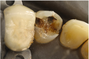
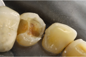
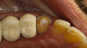
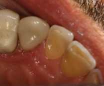
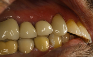
Summary – Tooth # 11 had large composite fillings with recurrent decay. The patient came with pieces of the composite in his hand. After doing the COAD (Clean Out And DIagnose and determining the restorability, we decided to restore the tooth with an Emax Crown using the CEREC CAD/CAM System. Our rationale for using Emax was due to the lack of interarch space due to wear. Our team felt that using a high strength monolithic material would not only provide better esthetics, but better use the space offered by the occlusal and axial reduction. To match this new crown with the natural looking canine on the other side of the arch (tooth #6), we utilized “Biogeneric reference” on the CEREC machine. The biogeneric reference function asked us if we wanted to mirror the contralateral tooth. Tooth #6 had a flat incisal surface, and the crown proposed by the CEREC came out with a normal architecture “triangular” shape. Minor modifications were made, but the patient wound up loving the computer’s design better than his existing tooth both in apperance and feel. It did raise an issue however that the proposal using biogeneric reference did NOT really resemble the tooth we wished to copy. This is probably why Biogeneric Copy using a diagnostic wax-up is still the preferred method to use for CEREC crowns made in the anterior region.
Decayed Canine
Tooth structure remaining after COAD procedure. Tooth remained Asymptomatic after Fuji Lining LC and definitive Core Flow build up was placed.
Prep
Final Occlusal View
Final Buccal view after occlusion was checked.

